Welcome to the new Gustave Roussy website
A new website designed to offer you a simpler, clearer, and more intuitive experience. Patients, caregivers, healthcare professionals, and donors: find information, news, and services more easily.
Imaging and Cytometry Platform
Joint Service Unit
US23 – AMMICa
Platform Type:
Technology Platform
Scientific Director:
Prof. Aline Renneville
Research Areas:
Spatial Analysis, Bioinformatics/Data Analysis, Cytometry, Biomedical Imaging, In Vivo/Preclinical Imaging, Cellular Imaging, Microscopy, Multiplex Imaging
Guardianship


Abstract
The PFIC is a shared service and development platform that serves as an interface between basic, translational, and clinical research programs.
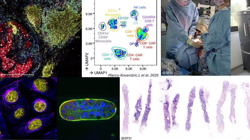
From sample analysis to data processing, it provides teams with expertise in cytometry (flow cytometry, mass cytometry, and cell sorting), bioinformatics, photonic microscopy, and in vivo imaging. Its area of expertise is specifically focused on cancer research, though it is open to other diseases as well. Its activities include providing technical and scientific support to internal and external academic teams as well as industry partners.
The platform participates in or leads R&D and technological innovation programs. It also provides theoretical and practical training—both locally and as part of national and international initiatives—to train young researchers and clinicians
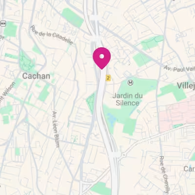
. Hours and Access: Research Pavilion No. 2, Ground Floor. Open
Access: For trained and independent users, certain platform equipment is accessible 24 hours a day, 7 days a week. For safety reasons, a “lone worker” form must be completed outside of staff hours.
Equipment reservations via the OpenIRIS platform.
Support and training: Our team is available Monday through Friday, from 9 a.m. to 6 p.m., to train you and assist you with the equipment, excluding holidays.
The PFIC is organized into three divisions:
• Cytometry Division: Operations Manager: Cyril Catelain; Scientific Advisor: Marie-Laure Arcangeli; Research Engineers: Philippe Rameau, Flavien Berthou; Bioinformatician: Alexia Alfaro.
• Microscopy Division: Operations Manager: Tudor Manoliu; Scientific Advisor: Guillaume Montagnac; Research Engineer: Flavien
Berthou• In Vivo Imaging Unit: Operations Manager: Muriel Abbaci, Scientific Advisor: Ingrid Breuskin, Research Engineer: Valérie Rouffiac

Services Offered
The Cytometry Center provides research units with state-of-the-art cytometers, analyzers, and sorters for conventional, spectral, and mass flow cytometry. It also offers the expertise of its staff for:
• Training and support in cytometry (flow, spectral, mass)
• Cell sorting (implementation and training)
• Mass imaging for molecular and cellular mapping of tissues •
Development of multiplexed panels (EasyPanel, Cytekcloud)
• Bioinformatics for unsupervised analysis of cytometry and imaging data (FlowJo, Kaluza, Omiq, Qupath)
The Cytometry Unit offers a bioinformatics service specializing in the analysis of multiparametric data.
The first component focuses on cytometry (flow cytometry, spectral cytometry, and mass cytometry) and concentrates on processing complex datasets using dimension reduction, clustering, and statistical analysis methods to identify biomarkers. In addition to analytical support (quality control, normalization, and correction for batch effects), the platform provides training, guiding users from beginner to expert levels to master the interpretation of these data.
A second component involves spatial proteomics using Hyperion technology. The platform has developed a dedicated analysis pipeline that enables the mapping of cellular interactions and the tumor microenvironment within tissues.
The Microscopy Center offers expert support for your projects, covering every step from sample preparation to data analysis.
• Sample Preparation: We provide training and guidance on advanced techniques such as immunofluorescence, Expansion Microscopy, and clearing methods.
• Microscopy Technologies: Access a diverse range of instruments, including wide-field, confocal, and multiphoton microscopy, as well as super-resolution techniques and holotomography microscopy (STED, FRET, TIRF, light-sheet microscopy, and spinning disk microscopy).
• Image Analysis: Training and personalized support for image data analysis software such as Fiji, Imaris, and Huygens.
• Expertise and Recognition: Accredited by IBISA and FBI France Bio Imaging, and members of the national networks RTMFM and GDR Imabio. Our team also contributes to the writing of scientific articles, ensuring cutting-edge expertise.
The In Vivo Imaging Center supports research teams in their preclinical imaging and in vivo cellular imaging projects. It provides expertise throughout the entire process, from protocol development to data analysis, ensuring compliance with European regulations.
Preclinical Imaging
• Training and support in in vivo imaging using bioluminescence, fluorescence, and microtomography (IVIS Lumina III/Spectrum, QuantumGX3).
• Longitudinal monitoring of preclinical tumor models, evaluation of the therapeutic efficacy of treatments, and validation of biomarkers under development.
In
vivo cellular imaging• High-resolution intravital imaging coupled with implantable optical devices or on exposed organs.
• Observation of tumor dynamics, cellular interactions, and therapeutic response.
• Developed through collaborative projects (oxidative stress, biodistribution, tissue regeneration, etc.).
The in vivo imaging division is part of the multidisciplinary SP2I (Surgery and Pathology Photonic Imaging) group, which is dedicated to the development and evaluation of photonic imaging technologies, from the cellular to the macroscopic scale, using in vivo and ex vivo protocols, to address clinical challenges in pathology and surgery.
SP2I is based on a collaboration between imaging specialists, surgeons, and pathologists. Its activities include the development of tools for tissue identification, margin assessment, functional evaluation, as well as diagnostic imaging and fluorescence-guided surgery in preclinical and clinical programs.
The group has enrolled more than 1,900 patients and collaborates with academic partners (PRISM, CREATIS, Polytechnique) and industry partners (Vivascope, SERB, etc.). It receives national and international funding (the League Against Cancer, Fondation Avenir, PHRC-K, BPI France, and the European Union).
Contact Us
If you are considering commissioning an analysis from us, please contact us to learn more about the available options, define the service, and obtain a quote: GD-PFIC@gustaveroussy.fr

Team
RAMEAU Philippe - Platform Scientific Director
Research Engineer or equivalent
Employer: Inserm
ROUFFIAC Valerie
IR - Research Engineer or equivalent
Employer: Gustave Roussy
CATELAIN Cyril - Platform Operations Manager
Research Engineer or equivalent
Employer: Gustave Roussy
Muriel ABBACI - Platform Operations Manager
Research Engineer or equivalent
Employer: Gustave Roussy
MANOLIU Tudor - Platform Operations Manager
Research Engineer or equivalent
Employer: Gustave Roussy
ALFARO Alexia
Design Engineer or equivalent
Employer: Gustave Roussy
BERTHOU Flavien
Research Engineer or equivalent
Employer: Gustave Roussy
ARCANGELI Marie-Laure - Scientific Manager, CR Platform
Research Associate and equivalent
positions Employer: Inserm
MONTAGNAC Guillaume - Platform Scientific Director
Research Director or equivalent
Employer: Inserm
BREUSKIN Ingrid - Platform Scientific Director
Hospital Practitioner
Employer: Gustave Roussy
Key Publications
Contact
- Address
-
Research Centers
20 Rue du Dr. Pinel
94 800 Villejuif