Two photon-counting CT scanners now equip Gustave Roussy's imaging department
Since September 2026, Gustave Roussy's Department of Medical Imaging has been equipped with a second photon-counting CT scanner, delivering higher image resolution while reducing the doses of X-rays and contrast agent administered to patients. This technology recently enabled the Institute to perform an iodine-free pulmonary CT angiogram, a world first.
Published on
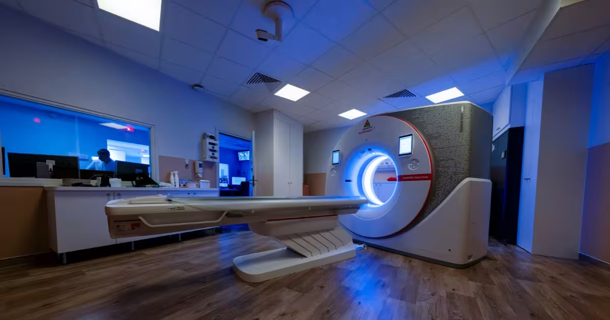
‘With the arrival of this second photon-counting CT scanner, we are taking another step forward: this technology is no longer reserved solely for paediatric patients and specialised examinations; it can now benefit a larger number of patients. For cancer imaging, this is an important advance, as it provides extremely precise images while reducing exposure to X-rays and, in some situations, the amount of contrast agent required’, says Professor Corinne Balleyguier, Head of Gustave Roussy's Department of Medical Imaging.
Since September 2026, Gustave Roussy has therefore had two photon-counting CT scanners among the three scanners on its imaging platform. This next-generation technology is transforming the way X-rays are detected and analysed:
- The NAEOTOM Alpha.Peak, the most powerful model in the range, designed for the most advanced applications and research.
- The NAEOTOM Alpha.Prime: the latest addition, a more accessible model based on the same photon-counting technology.
Unlike conventional CT scanners, these systems count individual photons and measure their energy, producing very high-resolution images for every examination.
Beyond the benefits already observed in clinical practice, Gustave Roussy's teams are continuing their work to explore the full potential of this technology in cancer care. Improved tumour characterisation, optimised treatment monitoring and the development of new diagnostic approaches are among the many applications currently under investigation.
This third scanner will also enable many examinations previously carried out outside Gustave Roussy to be brought back in-house, helping to streamline patient pathways at the Institute.
A world first already made possible by this new technology
The potential of this technology has already been demonstrated at the Institute. This summer, teams from the Department of Medical Imaging performed a pulmonary CT angiogram without iodinated contrast agent to diagnose a pulmonary embolism in a patient at risk of a severe allergic reaction to iodine.
The case began with a specific clinical situation. ‘An 81-year-old patient with breast cancer was referred to us as an emergency following the sudden onset of breathlessness. Doctors suspected a pulmonary embolism, a potentially serious complication that is usually diagnosed using CT angiography with an iodine-based contrast injection. However, the patient's medical record documented a severe allergic reaction to iodine during a previous examination’, recalls Dr Rémy Barbé, a radiologist at Gustave Roussy.
Faced with this situation, Gustave Roussy's Department of Medical Imaging chose a previously unreported approach, replacing the iodinated contrast agent with gadolinium, which is normally used for MRI examinations. This strategy was made possible by the photon-counting CT scanner, whose much greater sensitivity enables it to detect and make use of smaller quantities of contrast agent than a conventional CT scanner.
This world first was reported in a publication[1] in Diagnostic and Interventional Imaging in August 2026. It opens up a new diagnostic option for patients who, until now, could not undergo certain imaging examinations under optimal conditions, particularly because of a severe iodine allergy or renal impairment. This approach could therefore broaden diagnostic possibilities in many emergencies encountered in oncology, intensive care, emergency medicine, and surgery.
At Gustave Roussy, fundraising enables donors to contribute directly to the purchase of state-of-the-art medical equipment.
[1] Barbé, R., et al. Proof of feasibility of photon-counting CT pulmonary angiography using a gadolinium-based contrast agent for pulmonary embolism detection, Diagnostic and Interventional Imaging, 2026, ISSN 2211-5684, https://doi.org/10.1016/j.diii.2026.08.002.
Last news
-
Prevention and screening: getting ahead of lung cancer
To mark Thoracic Cancer Awareness Month and Tobacco-Free Month, two campaigns focusing on lung cancer, Gustave Roussy highlights its initiatives to advance prevention and screening among people at increased risk. Since its creation in 2021, the Institute’s Interception Lung programme has welcomed more than 900 participants, supporting them to stop smoking and offering personalised screening for the disease.
-
Gustave Roussy Meets His Audience
As part of its awareness-raising and fundraising efforts, Gustave Roussy is currently conducting a door-to-door outreach campaign in several towns in Val-de-Marne (Fresnes, Vitry-sur-Seine, Nogent-sur-Marne, Bry-sur-Marne, Le Kremlin-Bicêtre) from Monday through Saturday, 1:30 p.m. to 8:00 p.m.

-
Mens November: Take care of your jewelry its precious!
November is the month dedicated to raising awareness and promoting prevention of male cancers, which include prostate, testicular, and penile cancers. The goal is to raise public awareness of these diseases—which account for approximately 63,000 new cases in France each year—and to encourage men to perform testicular self-exams, a simple step that can help detect a lump, the nature of which must then be determined through further medical examinations. Gustave Roussy has extensive expertise in the treatment of male cancers, particularly through its Genitourinary Oncology Committee, which is deeply involved in research on these diseases.

-
Nuclear medicine: DART, a pioneering platform for the development of alpha therapy
Developed through a partnership between Gustave Roussy and the Paris-Saclay Cancer Cluster, the DART project aims to provide industry partners with a centre of excellence spanning preclinical, clinical and translational research to support the development of alpha therapy. This innovative nuclear medicine approach can destroy cancer cells more powerfully than standard approaches while reducing damage to surrounding healthy cells.