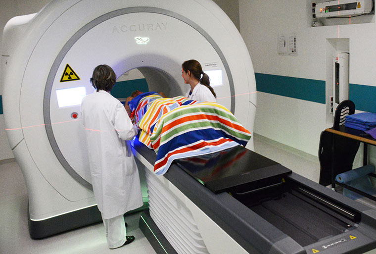
For high precision radiotherapy
Tomotherapy combines a CT scanning system with a radiotherapy machine. It is useful in the very precise treatment of tumours of complex shape which are located close to radiosensitive organs. It is particularly indicated in the treatment of large tumours such as certain sarcomas, poorly accessible pelvic tumours (prostate, uterine cervix, etc.) and ENT tumours.

At the beginning of each treatment session, a new CT image sequence is performed and targeting and dose of the irradiation are then adjusted to the findings.
Benefits
The integration of imaging capability and treatment delivery within the same apparatus results in:
- Individualisation of the treatments: each one is adjusted very precisely to the conformation of the tumour being treated, so as to minimise irradiation of non-tumour areas.
- The use of imaging during treatment: at each session CT images of the treated area are obtained, so that treatment can be guided with high precision by them.
The implication for patients is that treatment sequelae are very considerably reduced leading to a better quality of life. For the teams of doctors, physicists and staff handling patients and equipment, safety is increased by virtue of the system’s integration and precision.
Gustave Roussy has two tomotherapy machines.
